Error loading page.
Try refreshing the page. If that doesn't work, there may be a network issue, and you can use our self test page to see what's preventing the page from loading.
Learn more about possible network issues or contact support for more help.

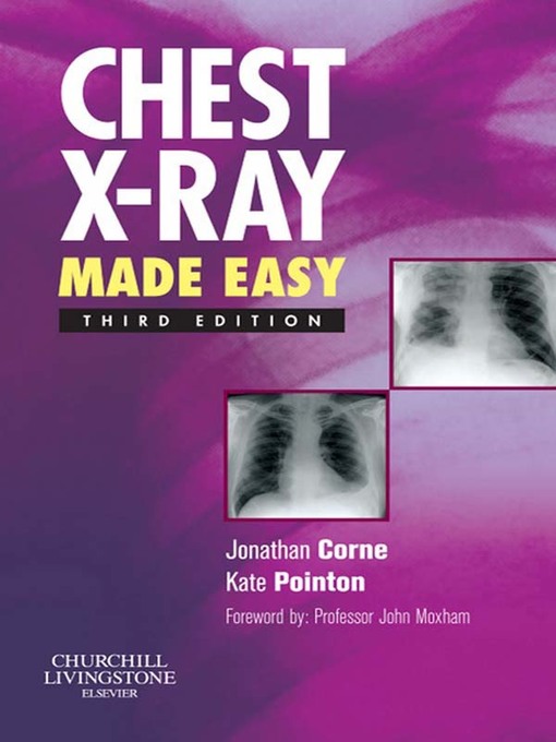
Chest X-Ray Made Easy E-Book

ebook
Translated into over a dozen languages, this book has been widely praised for making interpretation of the chest X-ray as simple as possible. It describes the range of conditions likely to be encountered on the wards and guides the doctor through the process of examining and interpreting the film based on the appearance of the abnormality shown. It then assists the doctor in determining the nature of the abnormality and points the clinician towards a possible differential diagnosis. It covers the common radiological problems the junior doctors are faced with starting with the appearance of the film, e.g. showing generalised shadowing or a coin lesion. It gives advice on how to examine an X-ray, how to check its technical quality and how to identify where the lesion is. All the X-rays are accompanied by a simple line diagram outlining where the abnormality is.
  • Covers the full range of common radiological problems.
  • Includes valuable advice on how to examine an X-ray.
  • Assists the doctor in determining the nature of the abnormality.
  • Points the clinician towards a possible differential diagnosis.
  • Now presented in two-colour to enhance the appearance of the text.
  • New material includes an introduction to thoracic CT scanning indicating the usefulness of these scans where appropriate.

  • Expand title description text
    Series: Made Easy Publisher: Elsevier Health Sciences Edition: 3

    OverDrive Read

    • ISBN: 9780702045097
    • File size: 3929 KB
    • Release date: October 1, 2009

    EPUB ebook

    • ISBN: 9780702045097
    • File size: 3929 KB
    • Release date: October 1, 2009

    Formats

    OverDrive Read
    EPUB ebook

    subjects

    Medical Nonfiction

    Languages

    English

    Translated into over a dozen languages, this book has been widely praised for making interpretation of the chest X-ray as simple as possible. It describes the range of conditions likely to be encountered on the wards and guides the doctor through the process of examining and interpreting the film based on the appearance of the abnormality shown. It then assists the doctor in determining the nature of the abnormality and points the clinician towards a possible differential diagnosis. It covers the common radiological problems the junior doctors are faced with starting with the appearance of the film, e.g. showing generalised shadowing or a coin lesion. It gives advice on how to examine an X-ray, how to check its technical quality and how to identify where the lesion is. All the X-rays are accompanied by a simple line diagram outlining where the abnormality is.
  • Covers the full range of common radiological problems.
  • Includes valuable advice on how to examine an X-ray.
  • Assists the doctor in determining the nature of the abnormality.
  • Points the clinician towards a possible differential diagnosis.
  • Now presented in two-colour to enhance the appearance of the text.
  • New material includes an introduction to thoracic CT scanning indicating the usefulness of these scans where appropriate.

  • Expand title description text